
Aortic and Great Vessel Imaging — Flashcards

Cardiac CT is done in _____ assisted by synchronous ECG gating.
The _____ sign is the acute angle formed at the edge of the false lumen in aortic dissection in axial cross-section
Cardiac CT is done in CAD and for _____ scoring.*scoring method?
_____ is a congenital heart disease characterized by a single large vessel arising from both ventricles before eventually splitting X-ray shows: "sitting duck heart" appearance
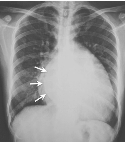
The x-ray given in the question shows the _____ sign suggestive of LA enlargement
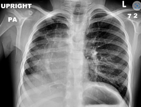
The X-ray shows _____ sign which is suggestive of partial anomalous pulmonary venous return.
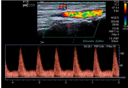
The given Doppler waveform is _____phasic, which is seen in severe stenosis of the artery:
_____ sign is seen in the appearance of splanchnic arteries in segmental arterial mediolysis (SAM).
Oreo cookie sign is seen in _____ on lateral view
Aortic and Great Vessel Imaging Flashcards for NEET-PG
Study 9 flashcards on Aortic and Great Vessel Imaging for NEET-PG Radiology. These active recall cards cover the key concepts, clinical associations, and high-yield facts from this chapter of Cardiovascular Radiology. Each card is designed to test your understanding rather than just recognition, building stronger and more durable memories for exam day.
For personalised spaced repetition scheduling and unlimited flashcards, download the Oncourse app.
Frequently Asked Questions
Are Aortic and Great Vessel Imaging flashcards free?
How many flashcards does this chapter have?
How should I use these flashcards for NEET-PG?
Are there more flashcards for Cardiovascular Radiology?
Want unlimited flashcards?
Get full access to all flashcards, spaced repetition, and progress tracking.
Start For Free