Central scotoma, Paracentral scotoma and temporal field defects can be tested by _____ocular visual field testing.
#2
Normal extent of the visual field: Temporal: _____Inferiorly: 70Nasal: 60Superiorly: 50
#3
Confrontation method is a type of _____ perimetry
#4
Lesions to the optic nerve result in _____-ocular and ipsi-lateral deficits in vision
#5
What pattern of visual loss is seen with LHON?_____
#6
Incongruous homonymous hemianopia, is usually seen with lesions of the _____
#7
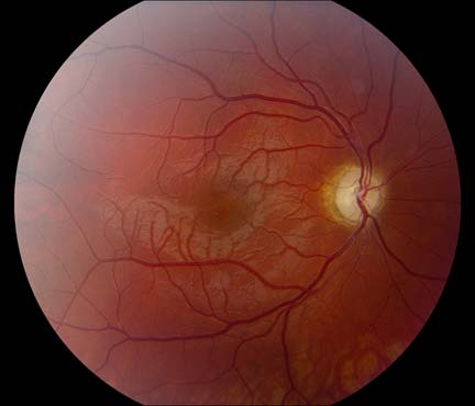
_____ is a condition of abnormal myelination of the nerve fibers of the retina, where myelination continues beyond lamina cribrosa and spreads into the retina beyond the optic disc.
#8
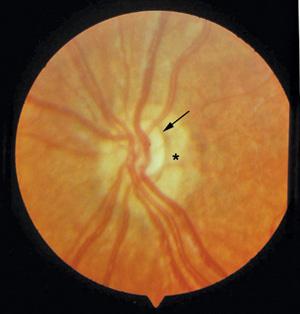
_____ sign is seen in optic nerve hypoplasia
#9
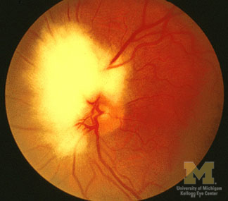
Hyperemic optic disc with telangiectatic (non-leaky on FFA) capillaries is suggestive of _____.
#10
Papilledema leads to blockage of _____, axonal swelling and collection of extracellular fluid
Visual Field Defects Indian Medical PG Flashcards - Medical Study Cards
Master Visual Field Defects with OnCourse flashcards. These spaced repetition flashcards are designed for medical students preparing for NEET PG, USMLE Step 1, USMLE Step 2, MBBS exams, and other medical licensing examinations.
Visual Field Defects Flashcard Deck - 10 Cards
Flashcard 1: Central scotoma, Paracentral scotoma and temporal field defects can be tested by _____ocular visual field testing.
Answer: mon
Flashcard 2: Normal extent of the visual field: Temporal: _____Inferiorly: 70Nasal: 60Superiorly: 50
Answer: 90/100
Flashcard 3: Confrontation method is a type of _____ perimetry
Answer: manual
Flashcard 4: Lesions to the optic nerve result in _____-ocular and ipsi-lateral deficits in vision
Answer: mon
Flashcard 5: What pattern of visual loss is seen with LHON?_____
Answer: Centrocecal scotoma
Flashcard 6: Incongruous homonymous hemianopia, is usually seen with lesions of the _____
Answer: optic tract
Flashcard 7: _____ is a condition of abnormal myelination of the nerve fibers of the retina, where myelination continues beyond lamina cribrosa and spreads into the retina beyond the optic disc.