_____is the most common complication of myopia, followed by rhegmatogenous retinal detachment.
#2
Indocyanin green angiography is used to visualize the _____ vasculature, especially in occult CNVM
#3
Diabetic retinopathy is more common in type _____ diabetes mellitus.
#4
_____ is useful in identifying cystoid macular edema and evaluating the vitreomacular interface.
#5
Best's disease is a form of macular dystrophy in which _____ accumulates in the central macula causing progressive central vision loss.
#6
Optical coherence tomography (OCT) in Best's disease reveals that the vitelliform material appears as a _____-shaped, hyperreflective, and homogenous lesion
#7
_____ would obscure the choroidal dye visualization in the corresponding area, but the retinal vessels overlying the area would, however, be visualized, on FFA.
#8
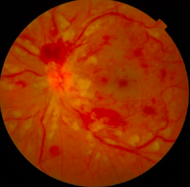
"Splashed sauce" appearance on ophthalmoscopy is seen in _____
#9
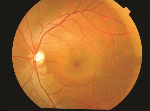
_____ is the spontaneous serous detachment of neurosensory retina in the macular region.
#10
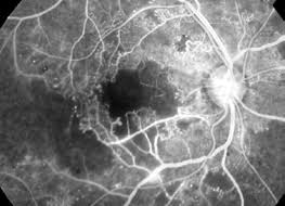
Increase in the size of the foveal avascular zone is seen in _____
Retinal Imaging Techniques Indian Medical PG Flashcards - Medical Study Cards
Master Retinal Imaging Techniques with OnCourse flashcards. These spaced repetition flashcards are designed for medical students preparing for NEET PG, USMLE Step 1, USMLE Step 2, MBBS exams, and other medical licensing examinations.
Flashcard 1: _____is the most common complication of myopia, followed by rhegmatogenous retinal detachment.
Answer: Macular degeneration
Flashcard 2: Indocyanin green angiography is used to visualize the _____ vasculature, especially in occult CNVM
Answer: choroidal
Flashcard 3: Diabetic retinopathy is more common in type _____ diabetes mellitus.
Answer: 1
Flashcard 4: _____ is useful in identifying cystoid macular edema and evaluating the vitreomacular interface.
Answer: OCT
Flashcard 5: Best's disease is a form of macular dystrophy in which _____ accumulates in the central macula causing progressive central vision loss.
Answer: lipofuscin
Flashcard 6: Optical coherence tomography (OCT) in Best's disease reveals that the vitelliform material appears as a _____-shaped, hyperreflective, and homogenous lesion
Answer: dome
Flashcard 7: _____ would obscure the choroidal dye visualization in the corresponding area, but the retinal vessels overlying the area would, however, be visualized, on FFA.
Answer: Submacular/subretinal hemorrhage
Flashcard 8: "Splashed sauce" appearance on ophthalmoscopy is seen in _____
Answer: CRVO
Flashcard 9: _____ is the spontaneous serous detachment of neurosensory retina in the macular region.
Answer: Central serous retinopathy
Flashcard 10: Increase in the size of the foveal avascular zone is seen in _____