
Diseases of the Retina — Flashcards

On this page
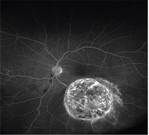
_____ can be seen on angiography in a patient of choroidal melanoma
In sympathetic ophthalmitis and Vogt-Koyanagi-Harada (VKH) syndrome, sub-retinal pigment epithelium (RPE) infiltrates corresponding to _____ nodules are seen
_____ would obscure the visualization of retinal vasculature and choroidal dye visualization on FFA.
_____ are cholesterol emboli that are seen in retinal arterioles in CRAO
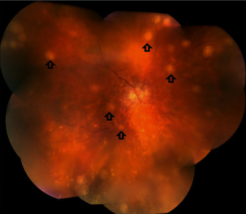
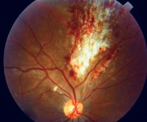
The following fundoscopy shows retinal _____ and white areas of retinal _____, classical presenting as "pizza pie retinopathy" (CMV)
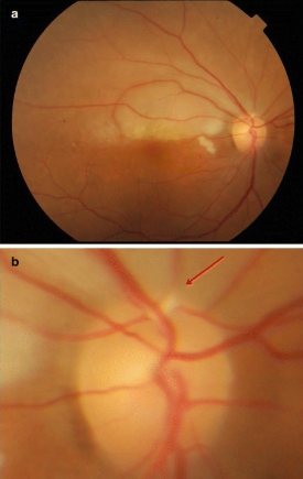
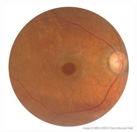
The given fundus finding is suggestive of a _____
_____ describes marked vascular sheathing that occurs in about 6% of patients with CMV retinitis
_____ would obscure the choroidal dye visualization in the corresponding area, but the retinal vessels overlying the area would, however, be visualized, on FFA.
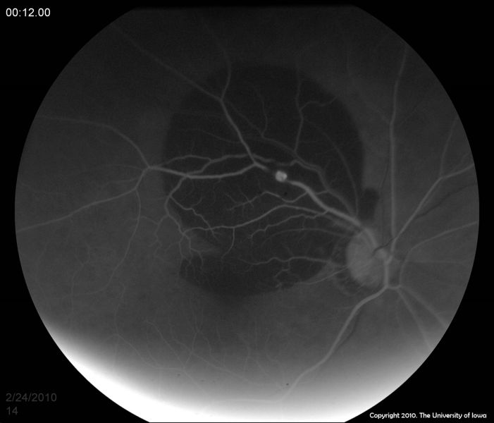
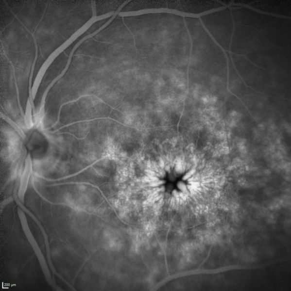
_____ would show the characteristic "Petaloid appearance" on FFA, due to the accumulation of dye in the outer plexiform layer at the fovea.
Dry macular degeneration is treated with _____ and antioxidant supplements; also smoking cessation if applicable
Study by Chapter
Retinal Anatomy and Physiology
Flashcards
Age-Related Macular Degeneration
Flashcards
Diabetic Retinopathy
Flashcards
Retinal Vascular Diseases
Flashcards
Retinal Detachment
Flashcards
Hereditary Retinal Dystrophies
Flashcards
Inflammatory Retinal Diseases
Flashcards
Retinal Tumors
Flashcards
Retinopathy of Prematurity
Flashcards
Retinal Imaging Techniques
Flashcards
Intravitreal Pharmacotherapy
Flashcards
Vitreoretinal Surgery
Flashcards
Want unlimited flashcards?
Get full access to all flashcards, spaced repetition, and progress tracking.
Start For Free