
Diseases of the Retina — Flashcards

On this page
"Splashed sauce" appearance on ophthalmoscopy is seen in _____
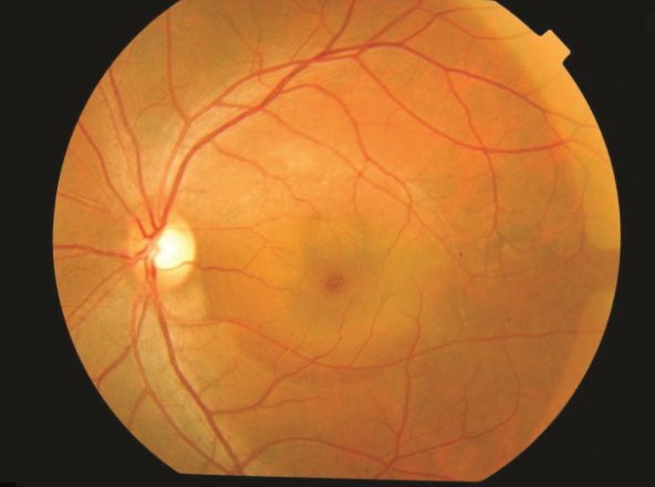
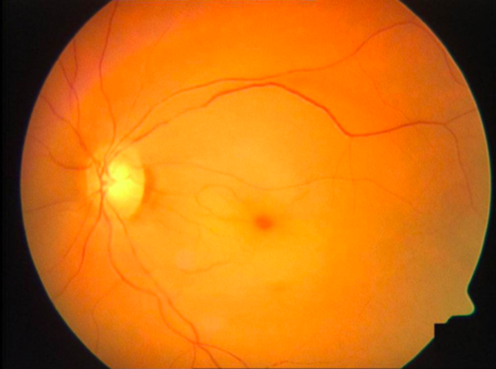
_____ is the spontaneous serous detachment of neurosensory retina in the macular region.
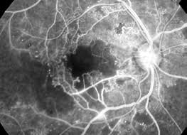
Increase in the size of the foveal avascular zone is seen in _____
_____ appearance of arteries and veins, and _____ like arteries are seen in CRAO (don't miss the obvious cherry red spot)
The image shows tobacco dust appearance (Shaffer's sign) suggestive of _____ retinal detatchment
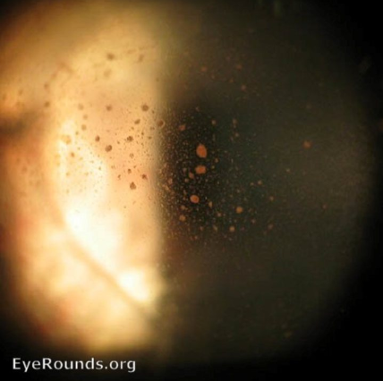
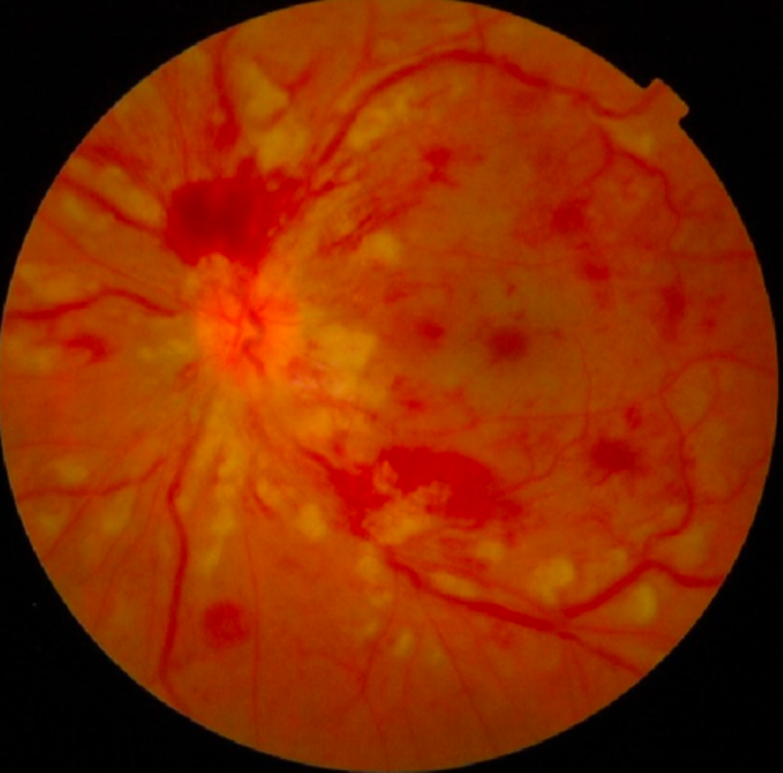
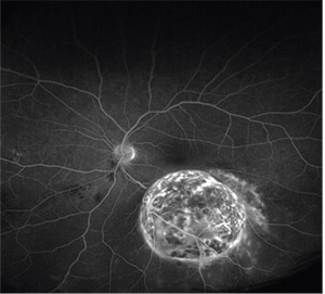
What pathology is shown in the given image? _____

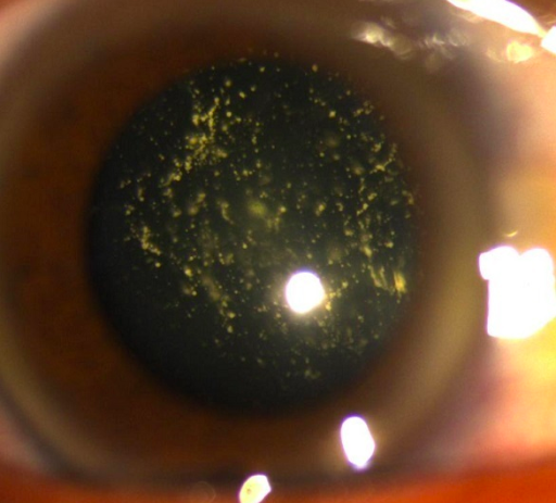
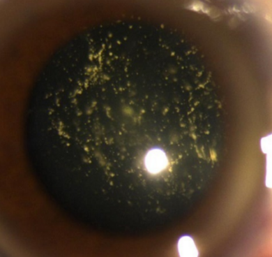
_____ are floaters, composed of calcium and lipids, seen in old age and in association with diabetes mellitus, hypertension, and high cholesterol levels.
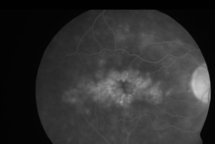
_____ edema occurs due to a break in the inner and outer blood-retinal barrier.
_____ has numerous yellowish-white vitreous opacities that don’t sediment when the eye is immobile
_____ can be seen on angiography in a patient of choroidal melanoma
Study by Chapter
Retinal Anatomy and Physiology
Flashcards
Age-Related Macular Degeneration
Flashcards
Diabetic Retinopathy
Flashcards
Retinal Vascular Diseases
Flashcards
Retinal Detachment
Flashcards
Hereditary Retinal Dystrophies
Flashcards
Inflammatory Retinal Diseases
Flashcards
Retinal Tumors
Flashcards
Retinopathy of Prematurity
Flashcards
Retinal Imaging Techniques
Flashcards
Intravitreal Pharmacotherapy
Flashcards
Vitreoretinal Surgery
Flashcards
About Diseases of the Retina Flashcards for NEET-PG
These Diseases of the Retina flashcards are designed for NEET-PG Ophthalmology preparation, using active recall to help you retain high-yield concepts, clinical correlations, and commonly tested facts. Each card prompts you to retrieve information from memory rather than passively reviewing notes, which research shows leads to significantly better exam performance.
The 215 cards in this deck cover the most important topics in Diseases of the Retina, including key mechanisms, diagnostic criteria, treatment protocols, and clinical pearls that frequently appear in NEET-PG examinations. Cards are organised by chapter so you can focus on specific areas or work through the entire topic systematically.
For personalised spaced repetition scheduling that adapts to your performance, unlimited flashcards, and detailed progress analytics, download the Oncourse app.
Frequently Asked Questions
Are Diseases of the Retina flashcards free on Oncourse?
How do flashcards help with NEET-PG preparation?
How many Diseases of the Retina flashcards are available?
Can I study Diseases of the Retina flashcards by chapter?
What topics do these Ophthalmology flashcards cover?
Want unlimited flashcards?
Get full access to all flashcards, spaced repetition, and progress tracking.
Start For Free