Internal thoracic artery is itself a branch of _____ artery
#2
Most of the joints in the thoracic cavity are _____ joints.
#3
The _____ nerve travels with the lateral thoracic artery through the axilla/lateral thorax
#4
The costal cartilages of _____ ribs form the subcostal angle.
#5
Which thoracic muscles are not pierced when doing pleural tapping in the midaxillary line?_____
#6
The arrangement of the components of the neurovascular bundle in the intercostal space from above downwards is _____.
#7
Vena caval hiatus is located at the level of _____, in the central tendon of diaphragm
#8
Which muscle layers are cut through while entering the pleural cavity for inserting an intercoastal drain?_____ and the intercostals.
#9
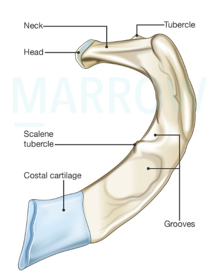
_____ passes anterior to the scalene tubercle of the 1st rib.
#10
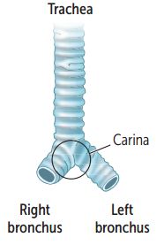
The carina is _____ to ascending aorta and _____ to descending aorta
Thoracic Wall and Diaphragm Indian Medical PG Flashcards - Medical Study Cards
Master Thoracic Wall and Diaphragm with OnCourse flashcards. These spaced repetition flashcards are designed for medical students preparing for NEET PG, USMLE Step 1, USMLE Step 2, MBBS exams, and other medical licensing examinations.
Thoracic Wall and Diaphragm Flashcard Deck - 10 Cards
Flashcard 1: Internal thoracic artery is itself a branch of _____ artery
Answer: subclavian
Flashcard 2: Most of the joints in the thoracic cavity are _____ joints.
Answer: plain synovial
Flashcard 3: The _____ nerve travels with the lateral thoracic artery through the axilla/lateral thorax
Answer: long thoracic
Flashcard 4: The costal cartilages of _____ ribs form the subcostal angle.
Answer: 7th, 8th, 9th, 10th ribs
Flashcard 5: Which thoracic muscles are not pierced when doing pleural tapping in the midaxillary line?_____
Answer: Transversus thoracis
Flashcard 6: The arrangement of the components of the neurovascular bundle in the intercostal space from above downwards is _____.
Answer: Vein-artery-nerve
Flashcard 7: Vena caval hiatus is located at the level of _____, in the central tendon of diaphragm
Answer: T8
Flashcard 8: Which muscle layers are cut through while entering the pleural cavity for inserting an intercoastal drain?_____ and the intercostals.
Answer: serratus anterior
Flashcard 9: _____ passes anterior to the scalene tubercle of the 1st rib.
Answer: Subclavian vein
Flashcard 10: The carina is _____ to ascending aorta and _____ to descending aorta
Answer: posterior
Keywords: Thoracic Wall and Diaphragm flashcards, medical flashcards, NEET PG preparation, USMLE Step 1 flashcards, Anki alternative, spaced repetition medical, OnCourse flashcards
Thoracic Wall and Diaphragm Flashcards for NEET-PG
Study 10 flashcards on Thoracic Wall and Diaphragm for NEET-PG Anatomy. These active recall cards cover the key concepts, clinical associations, and high-yield facts from this chapter of Thorax. Each card is designed to test your understanding rather than just recognition, building stronger and more durable memories for exam day.
For personalised spaced repetition scheduling and unlimited flashcards, download the Oncourse app.
Frequently Asked Questions
Are Thoracic Wall and Diaphragm flashcards free?
Yes, all flashcards on this page are completely free. You can study all 10 cards at no cost. For spaced repetition scheduling, download the Oncourse app.
How many flashcards does this chapter have?
There are 10 flashcards for Thoracic Wall and Diaphragm, covering the key concepts, clinical correlations, and high-yield facts from this chapter of Thorax.
How should I use these flashcards for NEET-PG?
Read through each prompt and try to answer before revealing the back of the card. This active recall approach builds stronger memories than passive reading. Combine flashcards with the study notes and MCQ practice for the best results.
Are there more flashcards for Thorax?
Yes. This page covers one chapter of Thorax. Visit the topic page to see all chapters and their flashcards under Anatomy.
Want unlimited flashcards?
Get full access to all flashcards, spaced repetition, and progress tracking.