INICET Image-Based Questions: Visual Recall, PYQ Review and Weak-Area Practice
Master INICET image-based questions with systematic visual recall strategies, PYQ analysis techniques, and targeted weak-area practice for higher scores.

INICET Image-Based Questions: Visual Recall, PYQ Review and Weak-Area Practice
You probably opened your INICET PYQ paper and froze at question 47. A chest X-ray with subtle shadows. Three ECG strips that looked identical. A histology slide where pink and purple merged into confusion.
You are not alone. Image-based questions now make up 25-30% of INICET — roughly 50-60 questions that can swing your rank by thousands. Unlike direct recall questions where you either know it or dont, image-based questions test pattern recognition, clinical correlation, and diagnostic reasoning under time pressure.
The difference between scoring 160 and 180+ on INICET often comes down to visual recall fluency. But most students approach image questions wrong: they memorize specific images instead of learning diagnostic patterns, skip systematic PYQ analysis, and never identify their true visual weak areas.
Here's how to turn image-based questions from your biggest weakness into your scoring advantage.
The INICET Image Question Reality Check
INICET 2026 follows a strict format: 200 questions across 4 blocks of 50, with 45 minutes per block and no backtracking. Within this structure, expect 50-60 image-based questions covering:
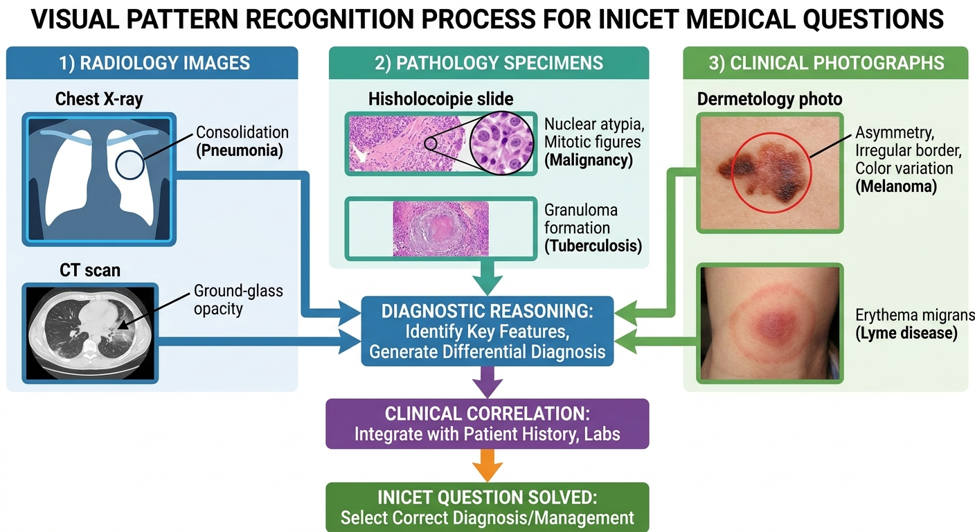
- Radiology (25-30% of images): Chest X-rays, CT scans, MRI, ultrasound
- Pathology (20-25%): Histology slides, gross specimens, special stains
- Clinical photographs (20-25%): Dermatology lesions, ophthalmology findings, ENT images
- ECG and cardiac (15-20%): Rhythm strips, echo findings
- Laboratory (10-15%): Blood smears, bone marrow, microscopy
The challenge isnt just recognition — its speed and systematic elimination under pressure.
Visual Recall vs Memorization: The Pattern Recognition Approach
Most students make a fundamental mistake: they try to memorize specific images rather than learning diagnostic patterns. This fails because INICET rarely repeats identical images. Instead, they test the same pathological pattern through different angles, stains, or imaging modalities.
Wrong approach: "This is the pneumonia X-ray from 2023 PYQ." Right approach: "This shows air-space consolidation in the right middle lobe — characteristic pattern includes increased opacity, air bronchograms, and preserved volume."The 3-Step Visual Analysis System
For every image question, force yourself through these three steps before looking at options:
Step 1: Modality and Orientation- What am I looking at? (X-ray, CT, histology, clinical photo)
- Normal anatomical landmarks
- Technical quality and limitations
- Primary abnormality (location, characteristics)
- Secondary changes or associations
- Differential diagnosis based on visual clues alone
- Read the clinical vignette
- Narrow differential based on history
- Select most likely diagnosis and next step
Building Your Visual Pattern Library
Create a systematic collection of diagnostic patterns rather than individual images. For each pattern, document:
- Core visual features: What makes this diagnosis recognizable?
- Variations: How does this pattern appear in different views or stains?
- Mimics: What other conditions look similar?
- Clinical correlation: How does the history help differentiate?
Strategic PYQ Review for Image Questions
Previous Year Questions remain the gold standard for INICET preparation, but image-based questions require specialized review techniques. Standard PYQ practice — attempt, check answer, move on — misses the deeper patterns.
The Cold Attempt Strategy
Before reviewing any explanations, attempt each PYQ paper under exam conditions: 180 minutes, no notes, no interruptions. For image questions specifically:
- Don't zoom in excessively (mirrors exam conditions)
- Time yourself per image (target 60-90 seconds max)
- Mark confidence level: "Certain," "Likely," "Eliminated to 2," "Pure guess"
Pattern Mapping Across Years
After cold attempts, map recurring image patterns across 2020-2025 INICET papers. You'll notice:
High-frequency patterns (appear 3+ times): Consolidation on chest X-ray, basic ECG rhythms, common skin lesions, standard histology patterns Medium-frequency patterns (appear 1-2 times): Complex radiology, rare pathology, specialty-specific images Single appearances: Often recent advances or one-off casesFocus 70% of your image practice on high-frequency patterns. These give maximum return on time investment.
The Diagnostic Feature Extraction Method
For every missed image question, document:
1. What you saw: Your initial impression and reasoning
2. What you missed: The diagnostic feature that led to the correct answer
3. Pattern abstract: How to recognize this pattern in future variations
4. Clinical hook: What clinical information should trigger consideration of this diagnosis
This builds a personalized atlas of your visual blind spots and their solutions.
Oncourse's Daily Plan feature schedules targeted image practice sessions based on your weak-area analysis, ensuring consistent exposure to problematic patterns without overwhelming your study schedule.
Weak-Area Identification for Visual Questions
Not all image-question mistakes are equal. Use systematic weak-area analysis to identify which visual domains need targeted practice.
The 4-Layer Mistake Classification
Layer 1: Recognition Level- Pattern recognition failure: Couldn't identify the basic abnormality
- Interpretation error: Saw the finding but misinterpreted significance
- Clinical correlation miss: Recognized pattern but failed to integrate with history
- Radiology: Which imaging modality and body system?
- Pathology: Gross specimens, histology, or special techniques?
- Clinical photos: Which specialty and anatomical region?
- Simple recognition: Single clear finding
- Complex integration: Multiple findings requiring synthesis
- Time pressure sensitivity: Accuracy drops when rushed
- High risk: Same error type across multiple similar images
- Medium risk: Occasional mistakes in specific contexts
- Low risk: One-off careless errors
Targeted Weak-Area Practice Blocks
Once you've identified patterns, create focused practice sessions:
Knowledge Gap Block: 20-30 images of the same pattern type (e.g., pneumonia chest X-rays) with systematic feature identification Speed Building Block: 15-20 mixed images with strict 45-second time limits to build rapid pattern recognition Integration Block: Complex cases requiring multiple-step reasoning, mimicking INICET's clinical vignette styleSchedule these blocks strategically. Practice your weakest visual domain when your concentration is highest, typically in morning study sessions.
Advanced Image Integration Strategies
Beyond basic pattern recognition, INICET tests your ability to integrate visual findings with clinical reasoning and next-step management.
The Clinical-Visual Bridge
High-scoring INICET questions often require connecting visual patterns to:
- Pathophysiology: Why does this pattern occur in this disease?
- Management implications: What does this finding mean for treatment?
- Prognostic significance: How does this change the patient's outlook?
Multi-Modal Pattern Recognition
INICET increasingly tests the same disease across different imaging modalities. A cardiac case might show:
- ECG changes (ST-elevation pattern)
- Chest X-ray findings (pulmonary edema)
- Echo abnormalities (wall motion)
- Lab correlation (troponin elevation)
Practice connecting these multi-modal patterns. When you see one component, actively predict what the other modalities should show.
Creating Visual Recall Loops for Long-Term Retention
Passive image viewing doesn't create lasting recall. You need active visual recall loops that strengthen pattern-diagnosis connections.
Active Recall Techniques for Images
Feature-First Recall: Look at an image, cover the question, and verbalize three diagnostic features before reading the clinical vignette. Progressive Revelation: Start with clinical history, predict what the image should show, then compare with actual findings. Cross-Modal Prediction: Given one type of image (e.g., chest X-ray), predict what other modalities (CT, echo) would show for this condition.Spaced Repetition for Visual Patterns
Unlike text-based facts, visual patterns need different spacing intervals:
- Initial learning: Daily exposure for pattern establishment
- Early consolidation: 3-day intervals for basic recognition
- Long-term retention: Weekly review of established patterns
- Pre-exam activation: Daily quick reviews of high-yield patterns
Integration with Broader INICET Preparation
Image-based question mastery shouldn't exist in isolation from your overall INICET strategy. Here's how to integrate visual recall practice with comprehensive preparation.
Subject-Wise Integration Approach
High-Yield Clinical Subjects (Medicine, Surgery, Pediatrics): Focus on diagnostic imaging and pathology correlation. Practice cases where clinical presentation connects to visual findings. Basic Science Subjects (Anatomy, Pathology, Pharmacology): Emphasize histology patterns and anatomical correlation imaging. Use visual mnemonics for drug mechanisms when possible. Low-Yield Subjects (Community Medicine, Forensic Medicine): Limit image practice to previously asked patterns. Don't over-invest time in uncommon visual questions from these areas.Mock Test Integration Strategy
In full-length mocks, track your image-question performance separately:
- Accuracy by image type: Which visual domains cost you marks?
- Time consumption: Are you spending too long on images?
- Confidence calibration: How often are you confident but wrong on visual questions?
When reviewing mocks, use Oncourse's Explanation Chat feature to clarify the diagnostic reasoning for missed image questions, particularly focusing on the visual features you should have recognized.
Exam-Day Visual Question Strategy
Your preparation culminates in exam-day execution. Image questions require specific tactical approaches during the actual INICET.
Block-by-Block Visual Strategy
Block 1-2 (Questions 1-100): Attempt image questions first within each block while your concentration is peak. Use full systematic analysis. Block 3-4 (Questions 101-200): Prioritize high-confidence image patterns. If you don't recognize a pattern within 30 seconds, eliminate obvious wrong answers and guess strategically.The 30-60-90 Rule for Images
- 30 seconds: Modality recognition and basic pattern identification
- 60 seconds: Clinical integration and differential narrowing
- 90 seconds: Final decision and moving on (never exceed this)
Strategic Elimination for Visual Questions
When pattern recognition fails, use systematic elimination:
1. Anatomical impossibility: Rules out options that don't match basic anatomy
2. Clinical mismatch: Eliminates diagnoses inconsistent with presented history
3. Pattern probability: Favors common presentations over rare variants
Even with limited pattern recognition, good elimination can achieve 60-70% accuracy on visual questions — enough to maintain competitive scoring.
Technology-Enhanced Visual Practice
Modern INICET preparation benefits from strategic technology use for image-based learning.
Digital Image Analysis Tools
Use software features that mirror exam conditions:
- Zoom limitations: Practice with standard screen resolution, avoid excessive magnification
- Brightness adjustment: Ensure comfortable viewing without eye strain during long practice sessions
- Multiple monitor setup: If available, practice switching between image and text like in CBT format
Creating Personal Visual Archives
Build curated collections of your problematic patterns:
High-Miss Images: Screenshots of consistently missed visual patterns with detailed analysis notes Speed Practice Set: 50-100 images for timed recognition drills Pre-Exam Review: Quick-access collection of high-yield patterns for final week reviewOncourse's "Find my weak areas" feature automatically identifies your most problematic visual patterns from practice performance, creating personalized focus areas for targeted improvement.
Measuring Progress and Adjusting Strategy
Track your visual recall improvement through quantified metrics rather than subjective feelings.
Key Performance Indicators
Pattern Recognition Speed: Time from image presentation to confident diagnosis Accuracy by Domain: Separate tracking for radiology, pathology, clinical photos Elimination Effectiveness: Success rate when using strategic guessing Time Allocation Balance: Percentage of total exam time spent on image questionsProgressive Difficulty Calibration
Structure your practice with increasing challenge levels:
Level 1: Clear, classic presentations with obvious diagnostic features Level 2: Atypical presentations requiring systematic analysis Level 3: Complex multi-modal cases with integrated reasoning Level 4: Time-pressured mixed practice simulating exam conditionsMove between levels based on consistent >80% accuracy at current level. Don't advance prematurely — solid foundation in basic patterns enables complex reasoning.
Weekly Assessment Protocol
Every week, take a standardized visual assessment:
- 25 mixed image questions
- 20-minute time limit
- Cover all major visual domains
- Track accuracy and pattern analysis
Use trends in weekly assessment to guide upcoming practice focus. Declining performance indicates fatigue or inadequate pattern consolidation.
Frequently Asked Questions
How many image-based questions should I practice daily?
Target 30-40 image questions daily: 20 from your identified weak areas and 10-20 mixed practice. Quality analysis matters more than quantity — better to thoroughly understand 30 images than superficially view 100.Should I create my own image flashcards?
Yes, but focus on diagnostic patterns rather than specific images. Create cards with key features, differential diagnosis points, and clinical correlation notes. Include images that show the same pattern in different presentations.How do I handle completely unfamiliar image patterns on exam day?
Use systematic elimination and clinical reasoning. Even without pattern recognition, you can often eliminate 2-3 options based on anatomical logic and clinical consistency, improving your guess probability significantly.What's the difference between INICET and NEET-PG image questions?
INICET emphasizes clinical correlation and next-step reasoning more than pure recognition. While NEET-PG might ask "What is the diagnosis?", INICET often asks "What is the next best step?" requiring integration of visual findings with management protocols.How important are recent imaging techniques and newer stains?
Focus 80% effort on classic, frequently tested patterns. Recent advances appear occasionally but shouldn't dominate your preparation. Master the fundamentals first, then add contemporary techniques if time permits.Can I compensate for weak visual recall with strong theoretical knowledge?
Partially, but not completely. Visual questions often test rapid pattern recognition under time pressure. Strong theory helps with differential diagnosis and clinical correlation, but you still need basic pattern recognition fluency to compete at top ranks.Prepare smarter with Oncourse AI — adaptive MCQs, spaced repetition, and AI explanations built for INICET success. Download free on Android and iOS.