Back
Types of Mechanical Injuries in Forensic Medicine: Abrasions, Contusions, Lacerations and Incised Wounds for NEET PG 2026
Master the 4 types of mechanical injuries in forensic medicine for NEET PG 2026: abrasions, contusions, lacerations, and incised wounds. Learn classification, medicolegal significance, and exam patterns.

Types of Mechanical Injuries in Forensic Medicine: Abrasions, Contusions, Lacerations and Incised Wounds for NEET PG 2026
You are staring at a forensic medicine MCQ. The question shows a wound image and asks you to classify it. Your mind races: "Is this an abrasion or a superficial laceration? Are those irregular edges typical of a contusion or laceration?"
Here's the thing — mechanical injury classification is pure pattern recognition. Master the 4 main types, their distinguishing features, and common NEET PG question patterns, and you'll never second-guess yourself again.
NEET PG forensic medicine has 8-12 questions annually. Around 40% focus on mechanical injuries. That's 3-5 marks sitting right there, waiting for students who know their wound types inside out.
Understanding Mechanical Injuries: The Foundation
Mechanical injuries result from physical force application to body tissues. They're the bread and butter of forensic medicine because every assault, accident, and suspicious death involves some form of mechanical trauma.
The classification is straightforward: blunt force trauma produces abrasions and contusions, while sharp force trauma creates lacerations and incised wounds. But here's where most students trip up — the devil is in the distinguishing features.
Why Classification Matters in Forensic Medicine
Wound classification isn't academic exercise. It determines:
Weapon identification — was it a knife, broken glass, or blunt object?
Force estimation — how much force was used?
Timeline reconstruction — when did the injury occur?
Legal implications — was it accidental, self-inflicted, or assault?
For mechanical injuries in forensic medicine, these distinctions can make or break a case.
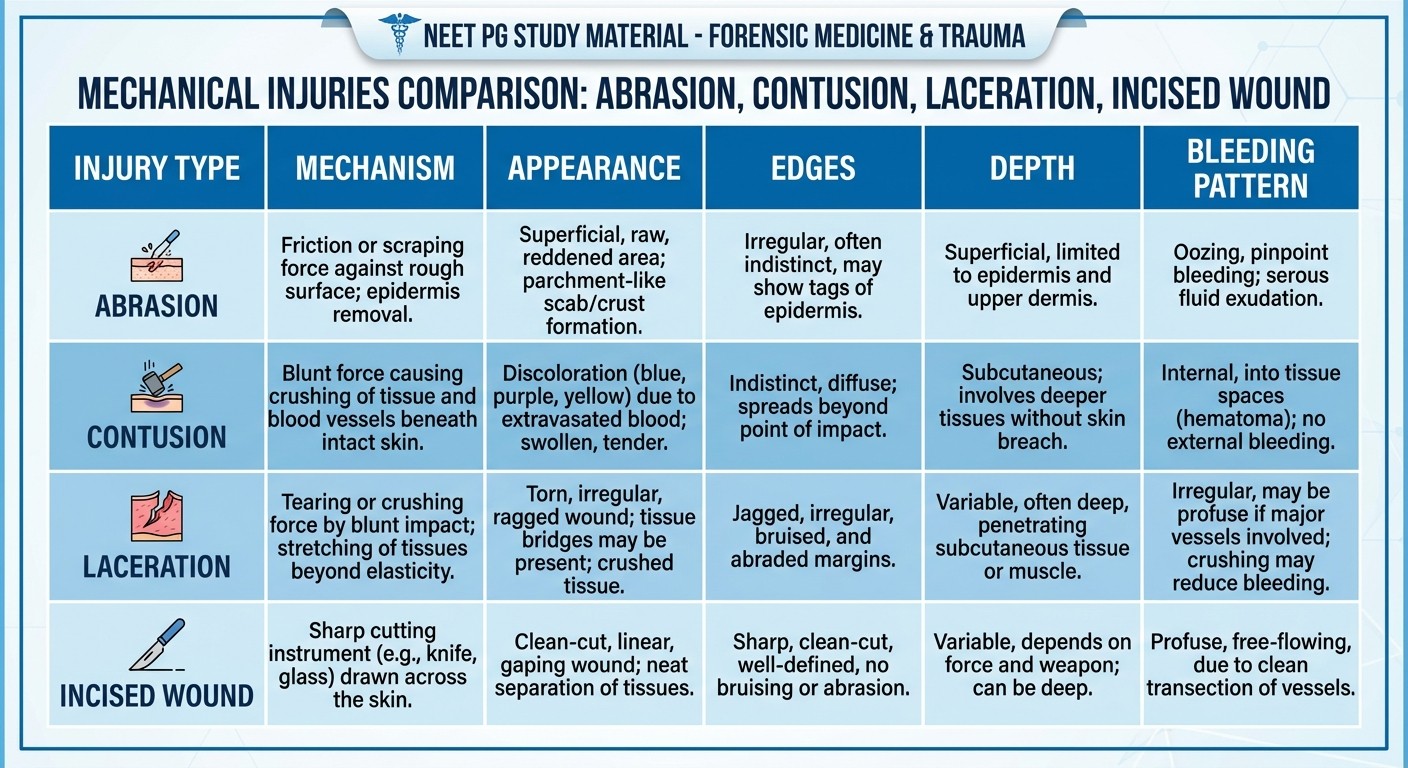
Type 1: Abrasions — The Scraping Injuries
Abrasions occur when skin slides against a rough surface, scraping away the superficial layers. Think road rash after a bike accident or rug burn from wrestling.
Key Features of Abrasions
Mechanism: Friction between skin and rough surface Appearance: Raw, weeping surface with exposed dermis Edges: Ill-defined, irregular borders Depth: Superficial — limited to epidermis and papillary dermis Bleeding: Minimal oozing, no active bleeding
Types of Abrasions Tested in NEET PG
1. Scratch abrasions — fingernail marks, animal claws
2. Graze abrasions — sliding contact with rough surface
3. Pressure abrasions — crushing against rough surface
4. Impact abrasions — sudden contact with textured object
The classic NEET PG trick question: "Which type of abrasion can indicate direction of force?" Answer: Graze abrasions show tissue tags pointing in the direction of movement.
Medicolegal Significance of Abrasions
Dating injuries: Fresh abrasions appear red and moist. After 8-12 hours, they form scabs
Pattern evidence: Can reproduce the shape of the causative object
Defense wounds: Fingernail scratches on attacker's hands or face
Sexual assault indicators: Abrasions around genitalia or inner thighs
For detailed abrasion patterns and healing timelines, check our blunt force injuries lessons.
Type 2: Contusions — The Bruising Injuries
Contusions result from blunt force rupturing small blood vessels under intact skin. The leaked blood creates the characteristic discoloration pattern.
Key Features of Contusions
Mechanism: Blunt trauma rupturing subcutaneous vessels Appearance: Discolored skin progressing through color stages Edges: Diffuse, gradually fading into normal skin Depth: Variable — from superficial to deep tissue involvement Bleeding: Internal bleeding with intact skin surface
Contusion Color Evolution Timeline
This is pure NEET PG gold. Questions love testing the color progression:
0-24 hours: Red to dark red
1-3 days: Purple to dark blue
4-6 days: Green shades appear
7-10 days: Yellow to brown
10-14 days: Fading to normal
Remember the mnemonic: "Red Purple Green Yellow Brown" — RPGYB.
Special Types of Contusions
Patterned contusions reproduce the shape of the weapon. A belt buckle leaves its distinctive pattern, tire treads create parallel lines, and knuckles produce grouped circular bruises. Aging of contusions helps establish timing in domestic violence cases. Multiple bruises in different color stages suggest repeated trauma over time.
NEET PG High-Yield Facts About Contusions
Contusions in dependent areas (back, buttocks) might be post-mortem hypostasis, not trauma
Deep contusions can occur without surface bruising in muscular individuals
Certain medications (anticoagulants, steroids) increase contusion severity
Location matters: Face and neck bruising is more significant than extremity bruising
Practice contusion identification with our forensic pathology questions.
Type 3: Lacerations — The Tearing Injuries
Lacerations are splits in skin and deeper tissues caused by blunt force trauma. The skin tears at its weakest points, creating irregular wound patterns.
Key Features of Lacerations
Mechanism: Blunt trauma causing tissue splitting Appearance: Irregular, gaping wound with tissue bridges Edges: Ragged, uneven, with crushing damage Depth: Variable — can extend to bone Bleeding: Moderate to severe depending on vessel involvement
Distinguishing Features of Lacerations
The key is recognizing tissue bridges — small strands of tissue spanning the wound. These occur because blunt trauma doesn't cleanly divide all tissue layers simultaneously.
Classic locations:
Scalp lacerations — skin splits against underlying skull
Shin lacerations — skin tears over the bony tibia
Eyebrow lacerations — common in blunt facial trauma
NEET PG Pearls for Lacerations
1. Tissue bridging distinguishes lacerations from incised wounds
2. Stellate patterns occur when blunt force strikes over bony prominences
3. Undermining of wound edges is common
4. Crushing artifacts at wound margins indicate blunt trauma
The classic exam scenario: "A 25-year-old presents with a 4cm wound over the right eyebrow with irregular edges and tissue bridges. What type of injury is this?" Answer: Laceration from blunt trauma.
Dive deeper into laceration patterns with our sharp force injuries lessons.
Type 4: Incised Wounds — The Cutting Injuries
Incised wounds result from sharp objects slicing through tissue. Think knife cuts, razor blade injuries, or glass lacerations.
Key Features of Incised Wounds
Mechanism: Sharp edge cutting through tissue Appearance: Clean, gaping wound with smooth edges Edges: Sharp, well-defined, without crushing Depth: Usually deeper at one end (tailing) Bleeding: Profuse bleeding from severed vessels
Characteristics That Distinguish Incised Wounds
No tissue bridges — this is the key differentiating feature from lacerations. Sharp objects cleanly divide all tissue layers. Tailing pattern — incised wounds are typically deeper at the beginning of the cut and shallow out toward the end. Clean edges without crushing or undermining distinguish them from blunt trauma injuries.
Types of Incised Wounds in NEET PG
1. Defense wounds — on hands and forearms of victims defending themselves
2. Hesitation marks — multiple superficial cuts before deeper fatal wound
3. Therapeutic cuts — surgical incisions or medical procedures
4. Self-inflicted cuts — typically on non-dominant arm, parallel orientation
Medicolegal Significance of Incised Wounds
Weapon characteristics can be determined from wound dimensions
Direction of force indicated by wound depth and tailing
Suicide vs homicide differentiation based on location and pattern
Self-defense evidence from distribution and characteristics
For comprehensive wound examination techniques, explore our injury examination lessons.
Differential Diagnosis: How to Tell Them Apart
Here's where NEET PG gets tricky. Questions often show borderline cases or ask you to differentiate between similar-looking injuries.
The Decision Tree Approach
Step 1: Is the skin surface intact?
Yes → Contusion
No → Proceed to Step 2
Step 2: Are the wound edges clean and sharp?
Yes → Incised wound
No → Proceed to Step 3
Step 3: Is the injury superficial (epidermis only)?
Yes → Abrasion
No → Laceration
Common NEET PG Confusion Points
Abrasion vs Superficial Laceration:
Abrasions don't penetrate full skin thickness
Superficial lacerations have tissue bridges
Laceration vs Incised Wound:
Lacerations have tissue bridges and irregular edges
Incised wounds have clean edges without bridging
Old Contusion vs Healing Abrasion:
Contusions progress through color changes
Abrasions form scabs and epithelialize
Practice Makes Perfect
Master these differentiations through targeted practice. Our mechanical injuries flashcards use spaced repetition to drill these distinctions until they're automatic.
NEET PG Question Patterns and High-Yield Topics
After analyzing years of NEET PG questions, certain patterns emerge consistently:
Most Frequently Tested Concepts
1. Wound classification from photographs (35% of questions)
2. Contusion color timing (25% of questions)
3. Distinguishing features between injury types (20% of questions)
4. Medicolegal significance of wound patterns (20% of questions)
Classic Question Formats
Pattern A: "A photograph shows a wound with [specific features]. What type of injury is this?" Pattern B: "A victim presents with bruising that is green-yellow in color. When did the injury likely occur?" Pattern C: "Which feature distinguishes a laceration from an incised wound?"
High-Yield Study Points
Tissue bridges are pathognomonic of lacerations
Contusion aging follows predictable color progression
Pattern wounds can identify specific weapons
Defense wounds indicate struggle and intent
Hesitation marks suggest suicidal intent
Medicolegal Documentation Requirements
Proper wound documentation is crucial for legal proceedings. NEET PG tests your knowledge of documentation standards.
Essential Documentation Elements
Wound Description:
Size (length, width, depth)
Shape and pattern
Edge characteristics
Associated findings
Location Documentation:
Anatomical landmarks
Distance from fixed points
Orientation description
Photographic Evidence:
Overall view with scale
Close-up details
Multiple angles if needed
According to the National Board of Examinations, forensic documentation must be detailed enough for legal testimony years later.
Age Estimation of Mechanical Injuries
Determining when an injury occurred is critical for timeline reconstruction in legal cases.
Healing Timeline for Each Injury Type
Abrasions:
0-6 hours: Fresh, oozing
6-24 hours: Scab formation begins
3-7 days: Complete scab coverage
1-2 weeks: Scab separation and healing
Contusions:
Use the RPGYB color progression timeline mentioned earlier
Lacerations:
0-24 hours: Fresh wound edges, active bleeding
3-5 days: Inflammatory response, edge swelling
1-2 weeks: Granulation tissue formation
2-4 weeks: Re-epithelialization complete
Incised Wounds:
Similar to lacerations but heal faster due to clean edges
Primary intention healing in 7-14 days
Factors Affecting Healing
Age — children heal faster than elderly
Nutrition — protein deficiency delays healing
Medications — steroids impair wound healing
Location — face heals faster than extremities
Infection — significantly delays healing process
Frequently Asked Questions
What's the most reliable way to distinguish between a laceration and incised wound?
The presence of tissue bridges is the gold standard. Lacerations from blunt trauma have tissue bridges spanning the wound because the force doesn't cleanly divide all layers. Incised wounds from sharp objects have no tissue bridges because the sharp edge cuts through all tissue uniformly.
How accurate is contusion color timing in real forensic cases?
Contusion aging has significant variability (±24-48 hours) and is affected by individual factors like age, skin color, medications, and location. While the color progression sequence is reliable, exact timing should be interpreted cautiously in legal contexts.
Can mechanical injuries occur post-mortem?
Yes, but they have different characteristics. Post-mortem injuries lack vital reaction (no inflammatory response, minimal bleeding, no healing). However, distinguishing ante-mortem from post-mortem injuries can be challenging and requires expert forensic evaluation.
What's the significance of defense wounds in assault cases?
Defense wounds typically appear on the palms, fingers, and forearms of victims trying to protect themselves. Their presence indicates active resistance and consciousness during the attack. The pattern and location help reconstruct the sequence of events.
How do you differentiate self-inflicted wounds from assault injuries?
Self-inflicted wounds are typically on accessible body areas (non-dominant forearm), have parallel orientation, include hesitation marks, and avoid vital areas initially. Assault injuries are more random in location, deeper, and often include defensive wounds.
What makes a wound "patterned" and why is it important?
Patterned wounds reproduce the shape or surface characteristics of the causative object. They're crucial because they can identify specific weapons, tools, or objects used in the assault, providing strong evidence linking a suspect to the crime.
Master these mechanical injury types and you'll handle any NEET PG forensic question with confidence. The key is recognizing patterns and understanding the physics behind each injury mechanism.
Prepare smarter with Oncourse AI — adaptive MCQs, spaced repetition, and AI explanations built for NEET PG. Download free on Android and iOS.