

💉 Core Concept - Clot E-vacuation
- Definition: Urgent removal of an occlusive thrombus or embolus from a vessel to restore perfusion, primarily for acute limb ischemia or massive PE.
- Goal: Prevent irreversible tissue damage (e.g., limb loss, organ failure).
- Main Approaches:
- Surgical (Open): Direct arteriotomy and clot extraction.
- Endovascular (Percutaneous): Catheter-directed mechanical thrombectomy (aspiration/fragmentation) or thrombolysis.
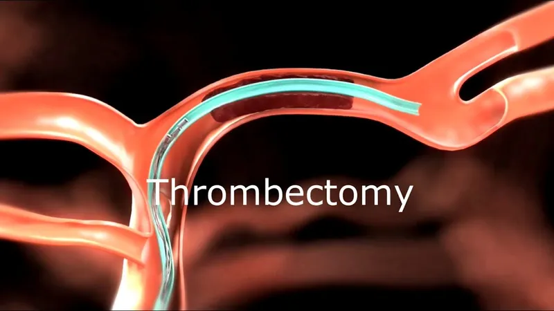
⭐ The Fogarty catheter is a classic tool for open embolectomy; its balloon tip is inflated past the clot and withdrawn, pulling the clot out.
🩸 Pathophysiology - The Blockage Buildup
-
Foundation: Virchow's Triad initiates thrombus formation.
- Endothelial Injury (e.g., atherosclerosis)
- Abnormal Blood Flow/Stasis (e.g., AFib, aneurysm)
- Hypercoagulability (e.g., Factor V Leiden, malignancy)
-
Sources of Occlusion:
- Embolism (~80%): Abrupt onset. Most common source is cardiac (AFib, post-MI thrombus).
- Thrombosis (~20%): Slower onset. Forms in situ on a pre-existing, ruptured atherosclerotic plaque.
⭐ Irreversible nerve damage begins after ~6 hours of ischemia, progressing to muscle necrosis. The "time is tissue" principle mandates urgent revascularization.
🛑 Diagnosis - Spotting the Stop Sign
-
Clinical Presentation: Suspect acute limb ischemia (ALI) with the classic 6 P's. 📌 Mnemonic: The 6 P's
- Pain (early, severe)
- Pallor
- Pulselessness (confirm with Doppler)
- Paresthesia (late sign)
- Paralysis (late sign)
- Poikilothermia (cool to touch)
-
Diagnostic Imaging:
- Bedside Doppler: Confirms absent arterial flow.
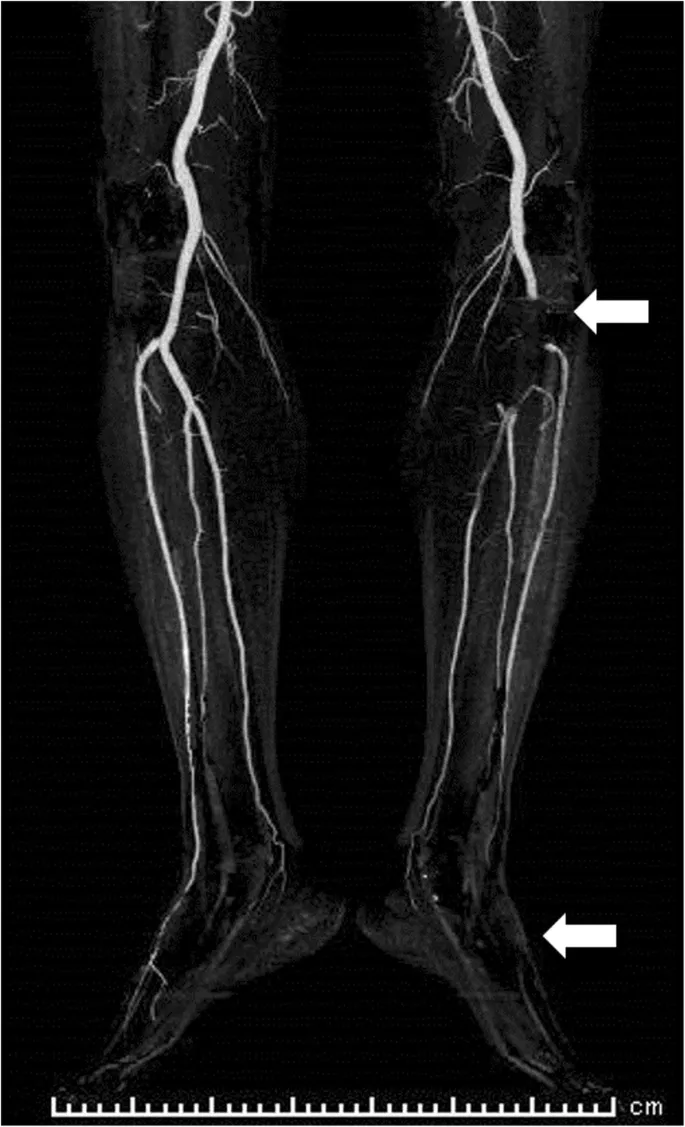
- CT Angiography (CTA): Gold standard. Rapidly identifies the location and extent of the occlusion ("the stop sign").
⭐ Time is tissue! Irreversible nerve damage and muscle necrosis can begin within 4-6 hours of ischemia. Paresthesia and paralysis are ominous signs indicating threatened limb viability.
🛠️ Management - The Extraction Mission
-
Goal: Rapidly restore perfusion to prevent irreversible tissue damage. Choice depends on limb viability (Rutherford class), clot location, and patient stability.
-
Catheter-Directed Thrombolysis (CDT):
- Low-dose thrombolytic (e.g., tPA) infused via a multi-side-hole catheter directly into the clot.
- Best for stable patients with viable/marginally threatened limbs (Rutherford I/IIa) and recent thrombus (<14 days).
- ⚠️ Risk: Hemorrhage (especially intracranial).
-
Percutaneous Mechanical Thrombectomy (PMT):
- Uses aspiration or rheolytic devices to remove/fragment the clot.
- Often adjunctive to CDT to reduce lytic dose and procedure time.
-
Surgical Embolectomy:
- Gold standard for immediately threatened limbs (Rutherford IIb).
- Involves arteriotomy and passage of a Fogarty balloon catheter.
⭐ Post-procedure, watch for reperfusion injury & compartment syndrome. Prophylactic fasciotomy may be needed, especially if ischemia > 4-6 hours.
💥 Complications - The Aftermath
- Reperfusion Injury: The most feared complication.
- Systemic "washout": Release of K+, lactate, myoglobin.
- Leads to: Hyperkalemia (↑$K^+$), metabolic acidosis, rhabdomyolysis → AKI.
- Local: Compartment syndrome from edema.
- Hemorrhage: Often due to aggressive anticoagulation or vessel trauma.
- Distal Embolization: Dislodged clot fragments occlude smaller, distal vessels.
- Re-thrombosis: Early failure at the embolectomy site.
⭐ Sudden reperfusion can cause a massive release of intracellular potassium, leading to life-threatening cardiac arrhythmias. Always have calcium gluconate ready.
⚡ High-Yield Points - Biggest Takeaways
- Primary Indication: Acute limb ischemia (ALI), classically presenting with the 6 P's (Pain, Pallor, Pulselessness, Paresthesia, Paralysis, Poikilothermia).
- Procedure: A Fogarty balloon catheter is passed beyond the clot, inflated, and withdrawn to mechanically extract the thrombus.
- Major Complication: Reperfusion injury, leading to compartment syndrome, rhabdomyolysis (↑CK), and life-threatening hyperkalemia.
- Post-Procedure: Immediate systemic anticoagulation (IV heparin) is crucial to prevent re-thrombosis.
- Adjunctive Surgery: Fasciotomy is often required to treat or prevent compartment syndrome after reperfusion.
Unlock the full lesson and continue reading
Signup to continue reading this lesson and unlimited access questions, flashcards, AI notes, and more