

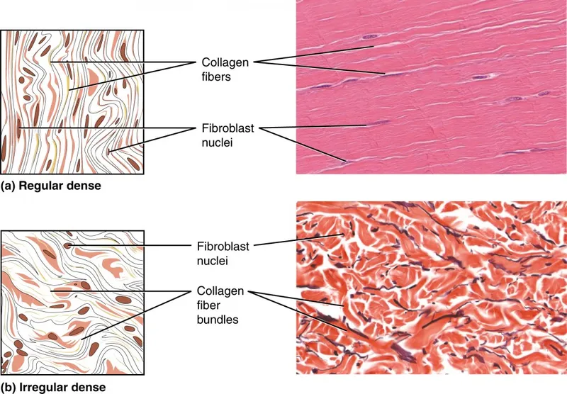
Dense Regular CT - The Body's Ropes
- Structure: Primarily parallel, densely packed Type I collagen fibers.
- Fibroblasts are the main cells, squeezed between fiber bundles.
- Very little ground substance and few blood vessels.
- Function: Provides exceptionally strong resistance to unidirectional tensile stress.
- Key Locations:
- Tendons (attach muscle to bone).
- Ligaments (attach bone to bone).
- Aponeuroses (flat, sheet-like tendons).
- 📌 Mnemonic: TLA
⭐ Exam Favorite: The avascular nature of dense regular CT is a critical concept. This poor blood supply is the primary reason why sprains (ligament injuries) and tendonitis heal so slowly and are prone to re-injury.
Dense Irregular & Elastic - Tough & Stretchy
-
Dense Irregular CT
- Structure: Primarily thick, irregularly arranged collagen fibers, with some elastic fibers and fibroblasts.
- Function: Withstands tension exerted in many directions; provides significant structural strength.
- Locations: Dermis of the skin, submucosa of the digestive tract, fibrous capsules of organs and joints.
-
Elastic CT
- Structure: Dense regular connective tissue dominated by elastic fibers, produced by fibroblasts.
- Function: Allows tissues to recoil after stretching; crucial for pulsatile blood flow and passive lung recoil.
- Locations: Walls of large arteries (aorta), ligamenta flava of the vertebrae, bronchial tubes.
⭐ Clinical Pearl: Marfan syndrome, a genetic disorder affecting fibrillin-1, critically weakens elastic tissues. This leads to life-threatening aortic aneurysms and dissections due to the loss of arterial wall recoil and strength.
Clinical Tie-Ins - When Fibers Fail
-
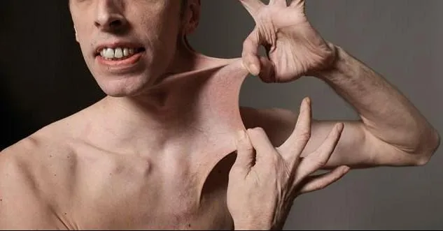
Ehlers-Danlos Syndrome (EDS): Defective collagen synthesis (e.g., Type V, Type III). Results in joint hypermobility, skin hyperextensibility, and easy bruising. Vascular type (Type III collagen defect) is life-threatening.
-
Osteogenesis Imperfecta (OI): "Brittle bone disease." Autosomal dominant defect in Type I collagen. Presents with multiple fractures, hearing loss, and dental imperfections.
- 📌 Mnemonic BITE: Bones (fractures), I (eye - blue sclerae), Teeth (dentinogenesis imperfecta), Ear (hearing loss).
-
Marfan Syndrome: Fibrillin-1 gene defect, not collagen. Affects elastic fibers. Leads to tall stature, arachnodactyly, lens dislocation, and aortic root dilation.
-
Scurvy: Vitamin C deficiency impairs collagen synthesis (hydroxylation step). Causes poor wound healing, bleeding gums, and perifollicular hemorrhage.
⭐ In Osteogenesis Imperfecta, the classic blue sclerae are due to thin scleral collagen revealing the underlying choroidal veins.
High‑Yield Points - ⚡ Biggest Takeaways
- Dominated by Type I collagen and fibroblasts, providing immense tensile strength to resist pulling forces.
- Dense regular tissue has parallel collagen fibers, ideal for structures like tendons and ligaments.
- The poor vascularization of dense regular CT is the primary reason for its notoriously slow healing time.
- Dense irregular tissue contains randomly arranged collagen bundles, resisting unpredictable stresses from multiple directions.
- Key locations for dense irregular include the reticular layer of the dermis, submucosa, and fibrous organ capsules.
Unlock the full lesson and continue reading
Signup to continue reading this lesson and unlimited access questions, flashcards, AI notes, and more